← Use Of Screens Effects And Mechanism Of Extracts Rich In Phenylpropanoids-polyacetylenes And Polysaccharides From Codonopsis Radix On Improving Scopolamine-induced Memory Impairment Of Mice →
Nanomechanical Mapping Of Three Dimensionally Printed Poly-ε-Caprolactone Single Microfibers At The Cell Scale For Bone Tissue Engineering Applications
This is one of the pictures featuring the Nanomechanical Mapping of Three Dimensionally Printed Poly-ε-Caprolactone Single Microfibers at the Cell Scale for Bone Tissue Engineering Applications. Many images associated with the Nanomechanical Mapping of Three Dimensionally Printed Poly-ε-Caprolactone Single Microfibers at the Cell Scale for Bone Tissue Engineering Applications can be utilized as your reference point. Below, you'll find some more pictures related to the Nanomechanical Mapping of Three Dimensionally Printed Poly-ε-Caprolactone Single Microfibers at the Cell Scale for Bone Tissue Engineering Applications.
 Title: Photographs of hollow microfibers and scaffold surfaces. a) hollow
Photographs of hollow microfibers and scaffold surfaces. a) hollow.
 Title: (pdf) nanomechanical mapping of three dimensionally printed poly-ε
(pdf) nanomechanical mapping of three dimensionally printed poly-ε.
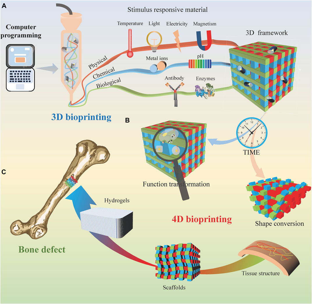
 Title: Frontiers | bioprinting for bone tissue engineering
Frontiers | bioprinting for bone tissue engineering.
