← Socket Shield Technique: A New Way To Deal With An Old Problem Recent Advances In Serum Biomarkers For Cardiological Risk Stratification And Insight Into The Cardiac Management Of The Patients With Hematological Malignancies Treated With Targeted Therapy →
Application Of Autofluorescence Microscopy And Laser Induced Fluorescence Methods To Study The Dynamics Of The Demineralization Primary Teeth Process In Vitro
Check out one of the pictures featuring the Application of autofluorescence microscopy and laser Induced fluorescence methods to study the dynamics of the demineralization primary teeth process in vitro. Many images associated with the Application of autofluorescence microscopy and laser Induced fluorescence methods to study the dynamics of the demineralization primary teeth process in vitro can be utilized as your reference point. Below, you'll find some additional pictures related to the Application of autofluorescence microscopy and laser Induced fluorescence methods to study the dynamics of the demineralization primary teeth process in vitro.
 Title: Representations of confocal laser scanning microscopy to visualize
Representations of confocal laser scanning microscopy to visualize. Confocal laser microscopy scanning fluorescence staining visualize representations fluorescent dichroic detector beam publication
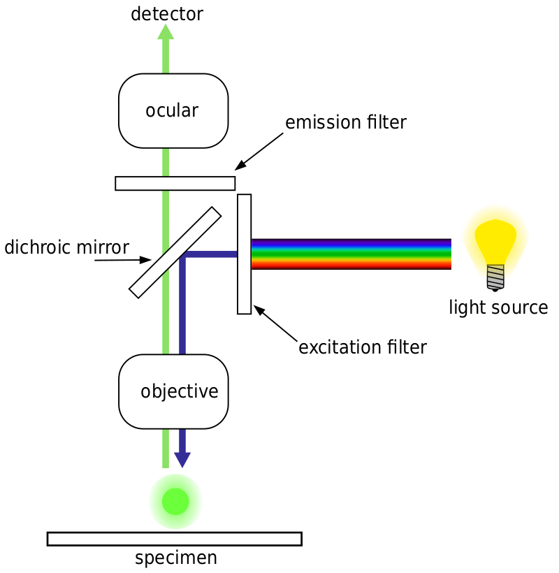
 Title: Fluorescence imaging
Fluorescence imaging. Fluorescence microscope microscopy epifluorescence confocal optical imaging epi excitation emission dichroic microscopes autofluorescence clipartbest acridine schematic homogenizers photometrics
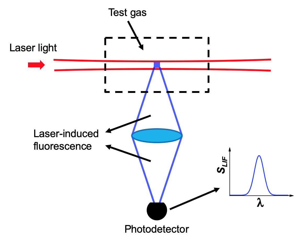
 Title: Laser-induced fluorescence | hanson research group
Laser-induced fluorescence | hanson research group.
